10+ diagram of replication fork
Is elongated away from the replication fork in segments. Replication Fork The replication fork is a region where a cells DNA double helix has been unwound and separated to create an area where DNA polymerases and the other enzymes.

A Schematic Representation Of The Replication Fork Polymerase Iii Download Scientific Diagram
Start studying Replication Fork Diagram.

. National Library of Medicine. Learn vocabulary terms and more with flashcards games and other study tools. Adenine A cytosine C.
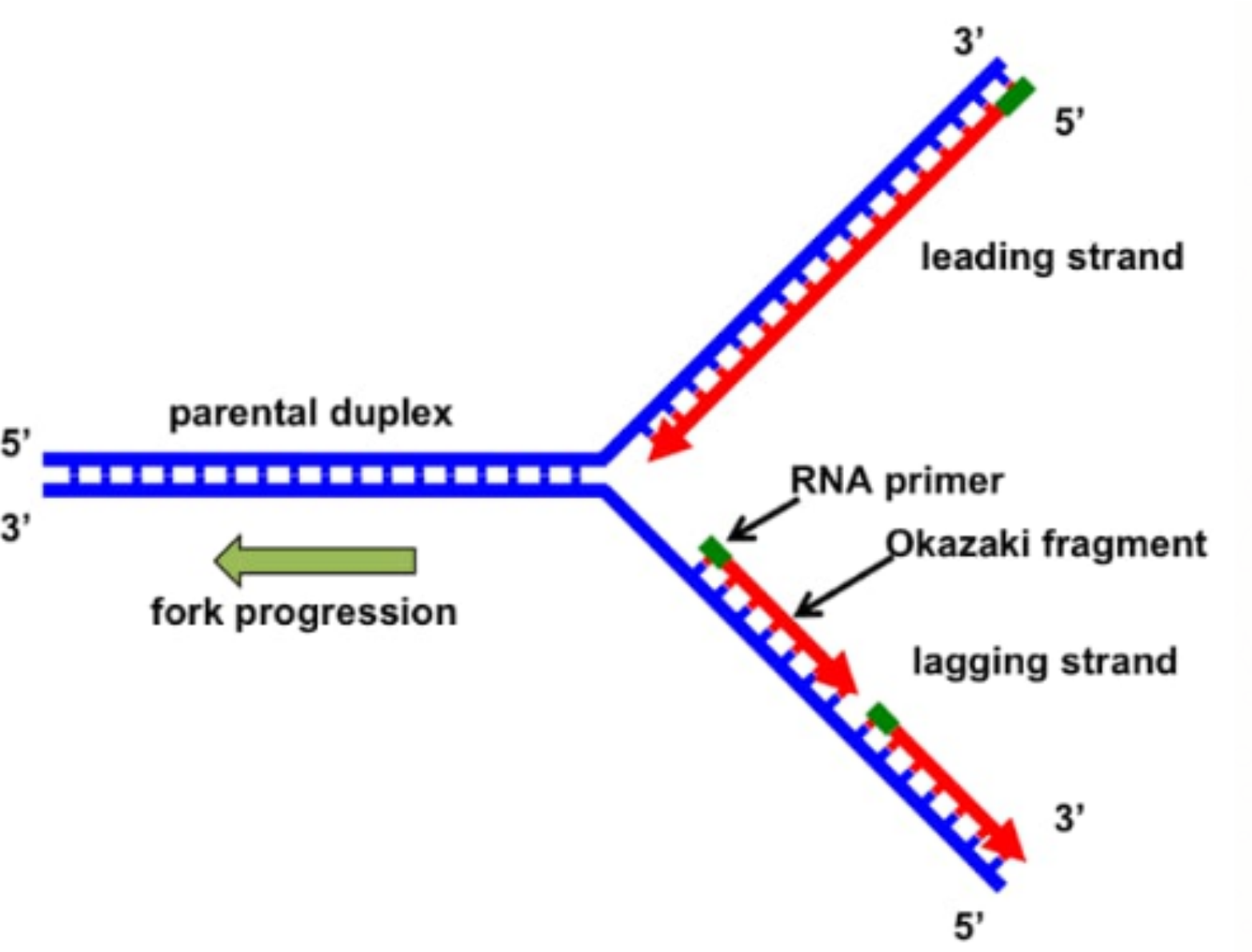
Replication Fork In the diagram can see one replication fork. There are two strands of DNA that are exposed. Synthesis of leading and lagging strands.
DNA replicates in a semi-conservative manner in which each individual strand is copied to form a new molecule of DNA. What is the purpose of a replication fork. This leads to two strands called leading and lagging strands.
The replication fork is a Y-shaped structure. The two strands can be labelled with isotopes using substrates that. Complement of the 5 to 3 parent strand.
Steps of DNA Replication Step 1. The replication fork is moving towards the left side of the diagram. The two strands can be labelled with isotopes using substrates that.
Strand that is being elongated discontinuously. Replicating fork is the structure of the DNA double helix after the unzipping by ligase enzyme. It forms at the repication bubble with the help of the enzyme DNA helicase.
The top strand will be used as a template to synthesize. The template for the synthesis of the lagging strand. DNA replicates in a semi-conservative manner in which each individual strand is copied to form a new molecule of DNA.
Explain why DNA replication is discontinuous on one strand. Formation of Replication Forks Before DNA can be reproduced it must first be unzipped into two single strands. In the following diagram of a replication fork which DNA strand a or b is a.
National Institutes of Health. The Function of the Replication Fork The replication fork is the area where the replication of DNA will actually take place. 1 mark Direction of fork movement 5 b.
DNA Replication Steps. 8600 Rockville Pike Bethesda MD 20894 USA. Diagram out the replication fork.
Why is eukaryotic DNA replication more complex than prokaryotic DNA replication.
Exactly Why In Dideoxy Dna Sequencing Do Dideoxynucleotides Halt The Replication Process Quora

Maintaining Genome Stability At The Replication Fork Nature Reviews Molecular Cell Biology
A Schematic Representation Of The Replication Fork Polymerase Iii Download Scientific Diagram

Eukaryotic Dna Replication Wikiwand

Draw A Labelled Diagram Of A Replicating Fork Showing The Polarity Why Does Dna Replication Occur Within Such Forks

Quantitative Assessment Of Rna Protein Interactions With High Throughput Sequencing Rna Affinity Profiling Abstract Europe Pmc

Simplified Model Of Replication Fork Download Scientific Diagram

Tolerance Of Deregulated G1 S Transcription Depends On Critical G1 S Regulon Genes To Prevent Catastrophic Genome Instability Sciencedirect

Oss Noise
A Draw A Labelled Diagram Of A Replicating Fork Showing The Polarity Why Does Dna Replication Occur Within Such Forks Sarthaks Econnect Largest Online Education Community

Our Blog Posts

Dna Replication Wikiwand
Replication Fork Y Fork Intermediate Molecular Biology

The Next Big Steps

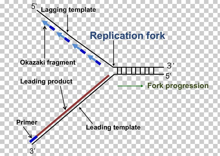
Dna Replication Replication Fork Enzyme Triangle Png Clipart Angle Diagram Dna Dna Replication Enzyme Free Png

Mink1 Regulates B Catenin Independent Wnt Signaling Via Prickle Phosphorylation Molecular And Cellular Biology
Phosphorylation Of The Mbf Repressor Yox1p By The Dna Replication Checkpoint Keeps The G1 S Cell Cycle Transcriptional Program Active Plos One